

Long Answer Questions - 5 Marks
Q1. Why are plants and animals made of different types of tissue?
Ans. Plants and animals are two different types of organisms. Plants are autotrophic organisms, so they prepare their own food by photosynthesis. Moreover, plants are stationary or fixed organisms. Since they do not consume or need much energy, so most of the tissues of plants are supportive. most of these tissues such as xylem, phloem, sclerenchyma and cork are dead tissues, i.e., they do not contain living protoplasm.
Animals on the other hand, are heterotrophic organisms. They have to move in search of food, mate and shelter; so they need more energy as compared to plants. Most of these tissues contain living protoplasm.
There are some tissues in plants which divide throughout life. They divide for the growth and reproduction of the plants. In contrast to plants, growth in animals is uniform.
Q2. Differentiate between parenchyma and collenchyma.
Ans.
|
Parenchyma |
Collenchyma |
|
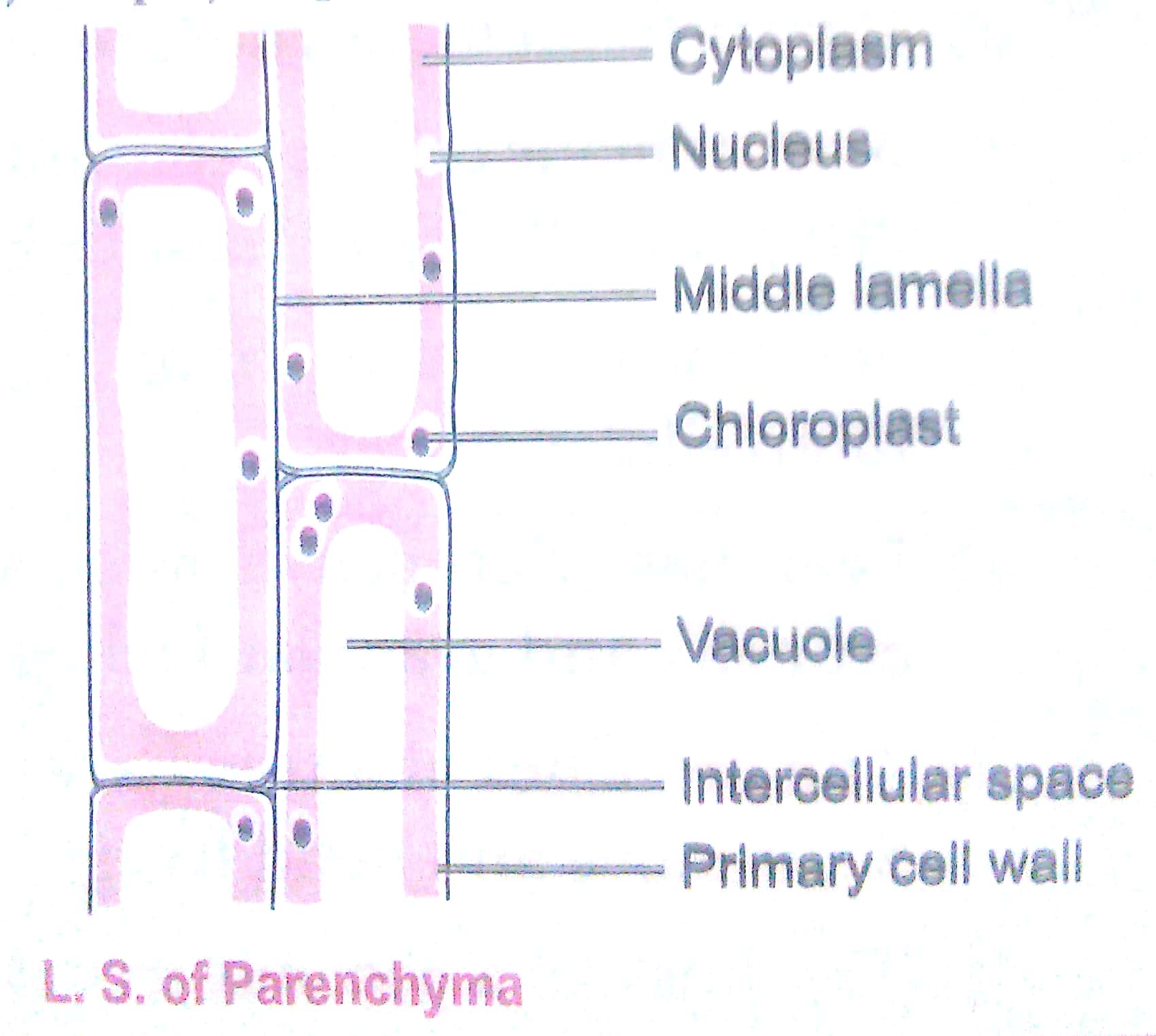
1. The tissue consists of thin-walled cells. 2. It is distributed in almost all the parts of the plant body. 3. The cells of parenchyma assimilate and store food. They also store waste products. 4. Parenchyma cells are loosely packed. |
1. The tissue consists of localised thickening in their cell walls. 2. It occurs mostly in the aerial parts of the plants restricted to the outer layers. 3. Collenchyma are the chief mechanical tissue of the young parts of the plant. 4. Collenchyma cells are compactly packed. |
Q3. Differentiate between collenchyma and sclerenchyma.
Ans.
|
Collenchyma |
Sclerenchyma |
|
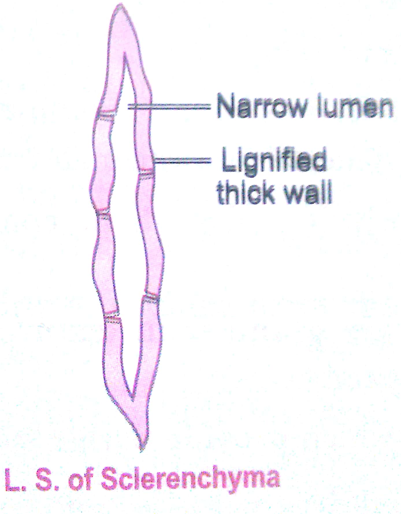
1. It consists of living cells. 2. The cells contain cytoplasm. 3. Its cell wall is cellulosic. 4. The thickening of cell wall is not uniform. 5. Lumen of cell is wide. 6. It provides mechanical support and elasticity to the plant body |
1. It consists of dead cells. 2. Cytoplasm is absent in these cells. 3. Its cell wall is lignified. 4. Cell wall thickening is uniform. 5. Lumen of the cell is narrow. 6. It is chiefly a mechanical tissue. |
Q.4. What is a neuron? Write the structure and functions of a neuron.
Ans. Nervous tissue contains highly specialised unit cells called nerve cells or neurons. Each neuron has the following three parts:
(i) The cyton or cell body: It contains a central nucleus and cytoplasm with characteristic deeply
stained particles, called Nissl granules.
(ii) The dendrites: These are short processes arising from the cyton.
(iii) The axon: It is a single, long, cylindrical process of uniform diameter. It carries impulses away from the cell body.
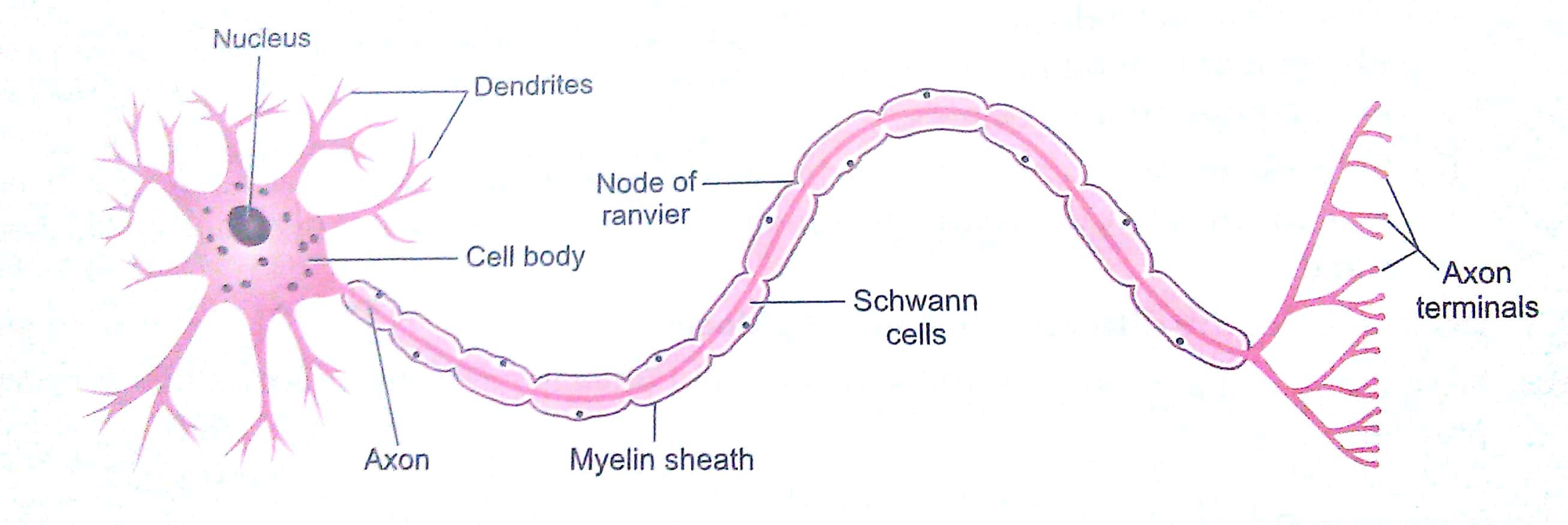
Functions:
Neurons have the ability to receive stimuli from within or outside the body and conduct impulses to different parts of the body. The impulses travel from one neuron to another neuron and finally to the brain or spinal cord.
Q. 5. Differentiate between meristematic tissue and permanent tissue.
Ans.
|
Meristematic tissue |
Permanent tissue |
|
1. The cells divide repeatedly. 2. The cells are undifferentiated. 3. The cells are small and isodiametric. 4. Intercellular spaces are generally absent. 5. Vacuoles are absent. 6. Metabolism occurs at high rate. 7. The cell walls are thin. |
1. The cells are derived from meristematic tissue and do not divide. 2. The cells are fully differentiated. 3. The cells are variable in shape and size. 4. Visible intercellular spaces are present. 5. Large vacuoles are present in mature cells. 6. Metabolism occurs at low rate. 7. Cell walls may be thin or thick. |
Q. 6. Briefly describe striated and smooth muscles with their functions.
Ans. The striated muscle fibres are long or elongated, non-tapering, cylindrical and unbranched. These cells have a number of nuclei called sarcolemma. These muscle fibres shows alternate dark and light stripes or striations and so they are called as striated muscles. These muscles occur in muscles of limbs, body wall, face, neck, etc.
Functions of striated muscles:
(i) Striated muscles are powerful and undergo rapid contraction and expansion.
(ii) Striated muscles provide the force for locomotion and all other voluntary movements of the body.
The smooth muscles are also known as unstriated or involuntary muscles. Smooth muscles occur as bundles or sheets of elongated fusiform or spindle-shaped cells or fibres. They are held together by loose connective tissue. These muscle fibres are uninucleate and do not bear any bands, stripes or striation across them.
These muscles are found in the walls of the alimentary canal and internal organs, ducts of glands and blood vessels. Smooth muscles are also found in the stomach, intestine, ureters, bronchi, iris of the eye, etc.
Functions of smooth muscles:
(i) Smooth muscles do not work according to our will, so they are also called involuntary muscles. Movement of food in the alimentary canal or the contraction and relaxation of blood vessels are involuntary movements.
(ii) smooth muscles contract slowly but can remain contracted for a long period of time. Due to this characteristic, the food passes to the next step of digestion in the alimentary canal.
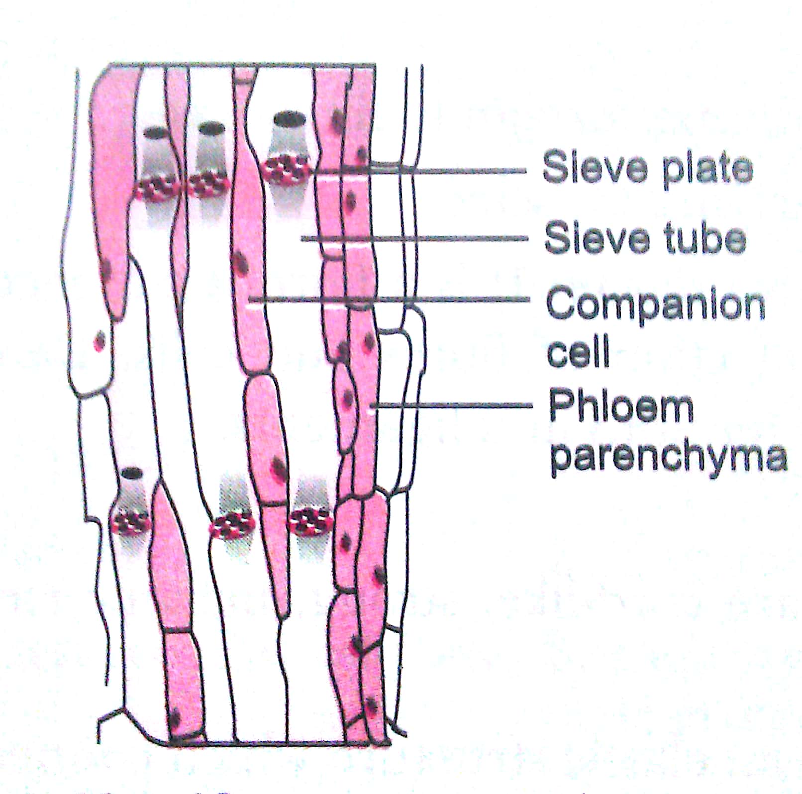
Q.7. Draw and identify different elements of phloem.
Ans. Sieve tubes, companion cells, phloem fibres and phloem parenchyma are the different elements of phloem.
Q8. What is a permanent tissue? Classify permanent tissue and describe them.
Ans. Permanent tissue are derived from meristematic tissue but their cells have lost power of division and have attained their definite forms.
Permanent tissue are classified into the following two types:
(i) Simple permanent tissue
(ii) Complex permanent tissue
(i) Simple Permanent tissue: These tissues are composed of cells which are structurally and functionally similar.
Simple permanent tissue are further classified into the following three types:
(a) Parenchyma: Parenchyma forms the bulk of the plant body. Parenchyma cells are living and possess the power of division.
(b) Collenchyma: Collenchyma tissue is also living. It is characterized by the deposition of extra cellulose at the corners of the cells.
(c) Sclerenchyma: Sclerenchyma cells are dead cells are dead cells and they are devoid of protoplasm. The cell walls of Sclerenchyma are largely thickened with deposition of lignin.
(ii) Complex permanent tissues: The complex tissue consist of more than one type of cells having a common origin. All these cells coordinate to perform a common function.
Complex tissue are of the following two types:
(a) Xylem: Xylem is a vascular and mechanical tissue. It is a conducting tissue. Xylem is composed of four different types of cells: (i) Tracheids (ii) Vessels (iii) Xylem parenchyma (iv) Xylem sclerenchyma.
Except xylem parenchyma, all other xylem elements are dead and bounded by thick lignified walls.
(b) Phloem: Like xylem, phloem is also vascular but has no mechanical function. Phloem is composed of following four element s: (i) Sieve tubes (ii) Companion cells (iii) Phloem parenchyma (iv) Phloem fibers.
Except phloem fibers, all other phloem elements are living.
Xylem and phloem are both conducting tissues and are also known as vascular tissues. Together, both of them constitute vascular bundle.
Q9. Describe the types of connective tissues along with their function.
Ans. There are five types of connective tissues:
(i) Areolar connective tissues: It is a loose and cellular connective tissue. It joins skin to muscles, fills space inside organs, and is found around muscles, blood vessels and nerves.
Functions:
(a) It acts as a supporting and packing tissue between organs laying in the body cavity.
(b) It helps in repair of tissues after an injury.
(c) It also helps in combating foreign toxins.
(d) It fixes skin to underlying muscles.
(ii) Dense regular connective tissue: It is a fibrous connective tissue, characterised by ordered and densely packed collection of fibres and cells. Dense regular connective tissue is the principal component of tendons and ligaments.
Functions:
(a) Tendons: Tendons are cord-like, strong, inelastic structures that join skeletal muscles to bones.
(b) Ligament: They are an elastic structure which connects bones to bones.
(iii) Adipose tissue: Adipose tissue is basically an aggregation of fat cells. The adipose tissue is abundant below the skin, between the internal organs and in the yellow bone marrow.
Functions:
(a) It serves as a fat reservoir.
(b) It provides shape to the limbs and the body.
(c) It keeps visceral organs in position.
(d) It forms shock-absorbing cushions around kidneys and eyeballs
(e) It acts as an insulator. Being a poor conductor of heat, it reduces heat loss from body, i.e., it regulates body temperature.
(iv) Skeletal tissue: The skeletal or supporting tissue includes bone and cartilage which form the endoskeleton of vertebrate body.
(a) Cartilage: The cartilage is a specialised connective tissue which is compact and less vascular. Cartilage can be found in ear pinna, nose tip, epiglottis, intervertebral discs, end of long bones, lower ends of ribs and rings of trachea.
(b) Bone: Bone is a strong and non-flexible tissue. Like cartilage, bone is also a specialized connective tissue.
Functions:
(a) Cartilage provides support and flexibility to the body parts. It smoothens the surface at joints.
(b) Bone provides shape and skeletal support to body.
(c) Bone protects vital body organs such as brain, lungs, etc.
(d) Bone anchors the muscles.
(v) Fluid connective tissue: Fluid connective tissue links the different parts of the body and maintains continuity in the body. It includes blood and lymph.
(a) Blood: In this tissue, cells move in a fluid or liquid matrix or medium called blood plasma. Blood occurs in blood vessels called arteries, veins, and capillaries which are connected together to form the circulatory system.
(b) Lymph: Lymph is a colourless fluid that has been filtered out of the blood capillaries,
Functions:
(a) Blood transports nutrients, hormones and vitamins to the tissues and transports excretory products from the tissues to the liver and kidney.
(b) Lymph transports the nutrients (oxygen, glucose) that may have filtered out of the blood capillaries back into the heart to be recirculated in the body.
(c) Lymph brings CO2 and nitrogenous wastes from tissues to the blood.
Q10. Differentiate between sclerenchyma and parenchyma tissues. Draw well labelled diagram.
Ans.
|
Sclerenchyma |
Parenchyma |
|
1. Cells are thick-walled and lignified. 2. Tissues are made up of dead cells. 3. No intercellular spaces between the cells are found. 4. Provides strength to the plant parts. 5. The cells are long and narrow, make the plant hard and stiff. The tissue is present in the stem around vascular bundles, in the veins of leaves and the hard covering of seed and nuts.
|
1. Cells are thin-walled and unspecialised. 2. These are living cells. 3. Cells are usually loosely packed with large intercellular space. 4. Stores nutrient and water in stem and roots. 5. Some cells contain chlorophyll called horenchyma and perform photosynthesis. Other cells have large air cavities called aerenchyma which provide buoyancy to the Hydrophytic plants.
|
Q11. Describe the structure and function of different types of epithelial tissues. Draw the diagram for each type of epithelial tissue.
Ans. Epithelial tissues are of following types:
(a) Simple squamous epithelium (b) Stratified squamous epithelium
(c) Columnar epithelium (d) Cuboidal epithelium
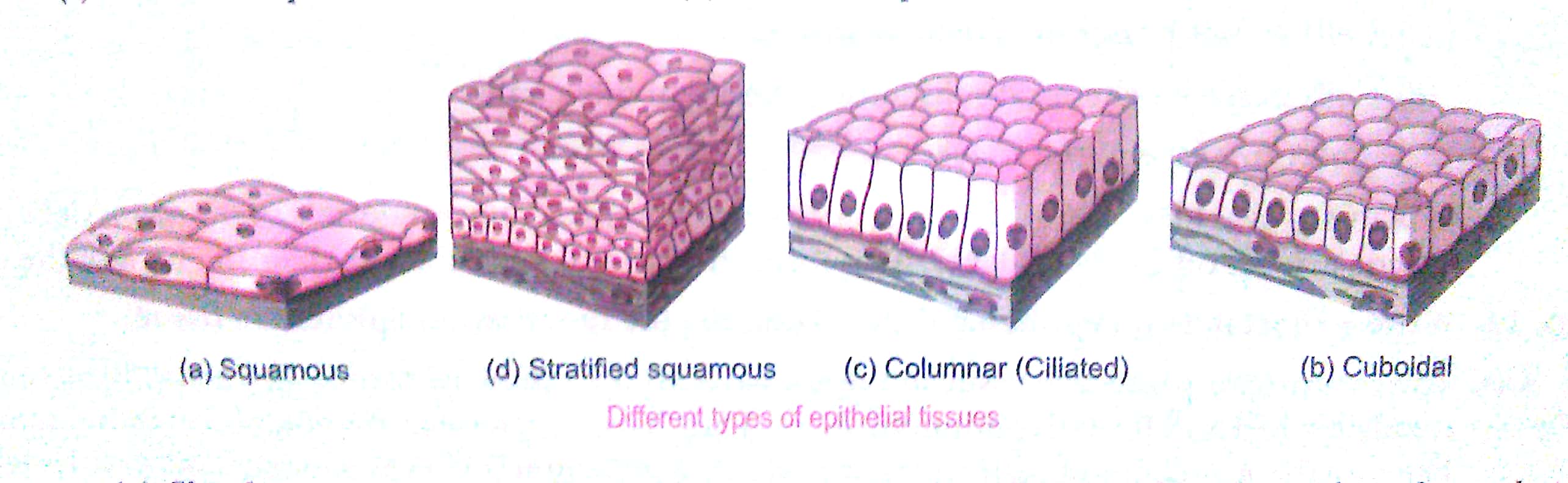
(a) Simple squamous epithelium: They are present in cells lining blood vessels or lung alveoli where transportation of substances occurs through a selectively permeable surface, there is a simple flat kind of epithelium.
(b) Stratified squamous epithelium: Skin epithelial cells are arranged in many layers to prevent wear and tear. Since, they are arranged in a pattern of layers, the epithelium is called stratified squamous epithelium.
(c) Columnar epithelium: Where absorption and secretion occur, as in the inner lining of the intestine, these tall epithelial cells are present. This columnar epithelial facilitates movement across the epithelial barrier. In the respiratory tract, the columnar epithelial tissue also has cilia, which are hair-like projections on the outer surfaces of epithelial cells. These cilia can move and their movement pushes the mucus forward to clear it. This type of epithelium is thus ciliated columnar epithelium.
(d) Cuboidal epithelium: These form the lining of the kidney tubules and ducts of salivary glands where these provide mechanical support. Sometimes, a portion of the epithelial tissue folds inward and a multicellular gland is formed. This is glandular epithelium.
Q12. Give reasons:
(a) Meristematic cells have a prominent nucleus and dense cytoplasm but they lack vacuole,
(b) Intercellular spaces are absent in sclerenchymatous tissues.
(c) We get a crunchy and granular feeling when we chew pear fruit.
(d) Branches of a tree move and bend freely in high wind velocity.
(e) It is difficult to pull out the husk of a coconut tree.
Ans. (a) Meristematic cells are continuously dividing cells so they have a prominent nucleus and dense cytoplasm. But since these cells do not store food material or waste materials, they lack vacuole.
(b) Sclerenchyma cells have lignified cell walls which makes them compact and leaves no intercellular spaces.
(c) Pear has sclerenchymatous stone cells which are granular in texture. Hence, we get the crunchy and granular feeling while chewing a pear.
(d) The branches of a tree have collenchyma cells which provide tensile strength to plant parts. So, it move and bend freely when wind blows.
(e) The husk of a coconut tree is made up of sclerenchyma cells which have lignified cell walls. Lignin makes the cells compact and leaves no intercellular spaces.
Q13. List the characteristics of cork. How are they formed?
Ans. The characteristics of cork are as follows
(a) Cells of cork are dead at maturity
(b) These cells are compactly arranged
(c) Cells do not posses intercellular spaces.
(d) Cells possess a chemical substance 'suberin' in their walls.
(e) There are several thick layers.
As plants grow older, a strip of secondary meristem replaces the epidermis of the stem. Cells on the outside are cut off from this layer, This forms the several-layer thick cork or the bark of the tree.
Q14. Write a short note on epithelial tissue. Describe the functions of epithelium tissue.
Ans. The covering or protective tissue in the animal body are epithelial tissues. Epithelial tissue cells are tightly packed and form a continuous sheet. They have only a small amount of cementing material between them and almost no intercellular spaces. Epithelium covers most organs and cavities within the body. It forms a barrier to keep different body systems separate. The skin, the lining of the mouth, the lining of blood vessels, lung alveoli and kidney tubules are all made of epithelial tissue.
Functions of epithelial tissue:
(i) Epithelial cells protect the underlying cells from drying, injury and chemical effects. protect the body from viral or bacterial infections.
(ii) It helps in the absorption of water and nutrients.
(iii) It performs secretary function by secreting useful chemicals like sweat, saliva, enzymes from the food, etc., in the body.